White
Type: Polygenic
Issues: N/A
First Produced In: Unknown
Availability: Average
Last Updated: 2022-04-26
Do you have any suggestions or corrections for this article?
Click here to contribute feedback
About
About white pigmentation:
Chromatophores are pigment-containing and light-reflecting cells found in amphibians, fish, reptiles, crustaceans, and cephalopods. They are largely responsible for generating skin and eye colour in cold-blooded animals and are generated in the neural crest during embryonic development.
The white pigmentation found in crested geckos is most likely caused by a lack of melanophores.
History
No history yet
Appearance
Body
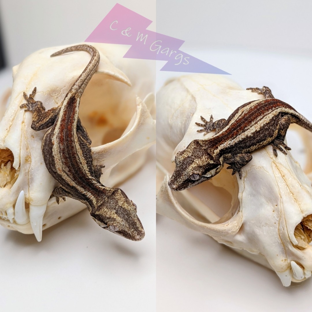
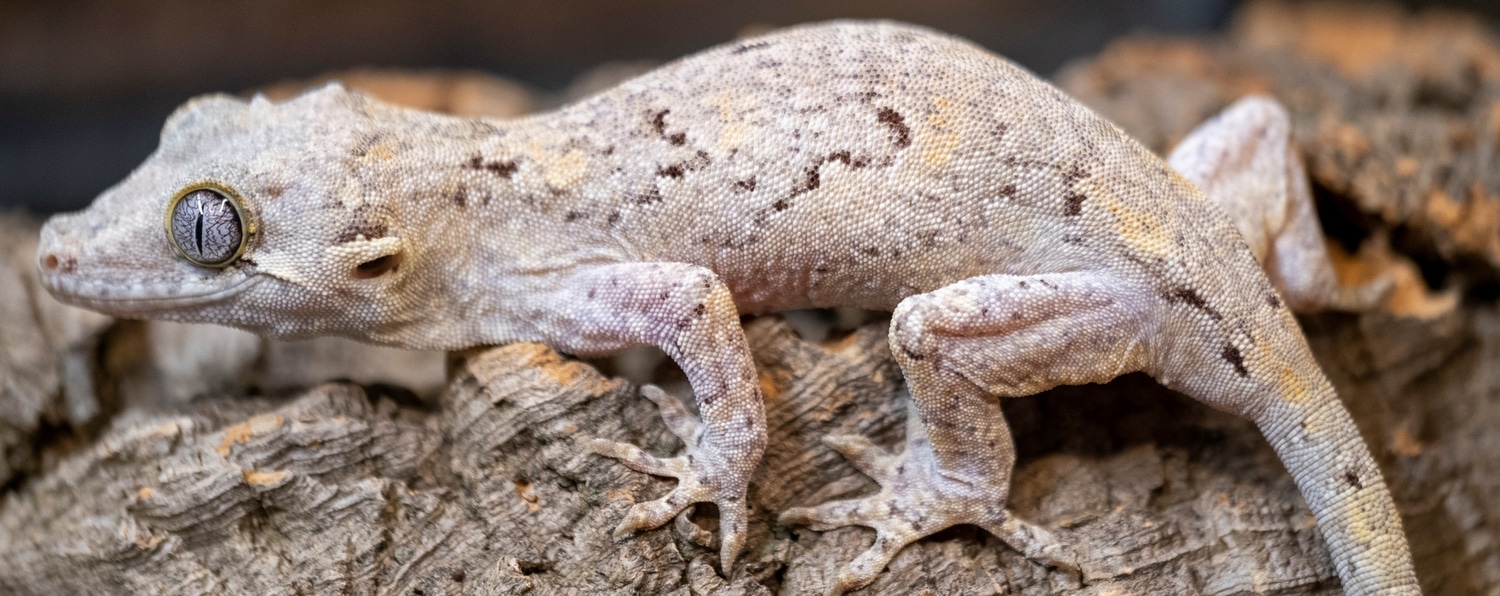
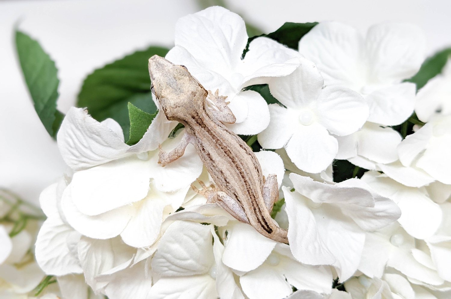
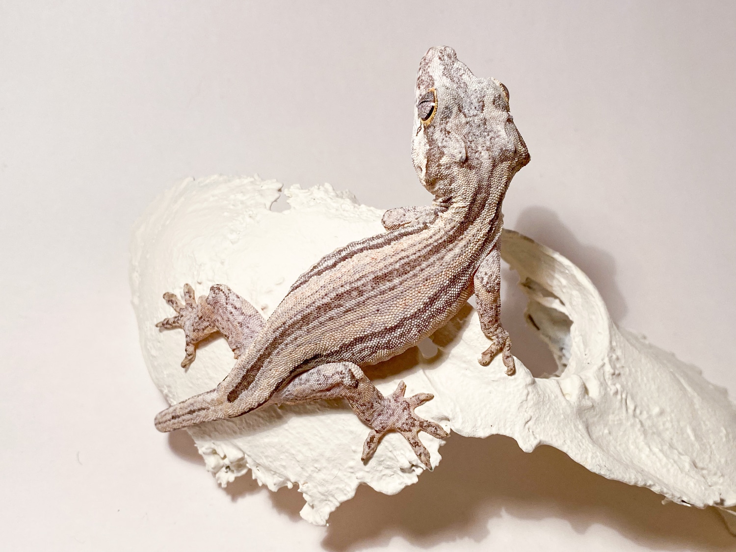
Generally only seen on a striped gecko (though there is an exception), this color pattern isn’t as common as other colors and is best visible when the gecko is fired up. In a dark fired gecko, the white stripe is often the side bar, and should be relatively solid. In young geckos exhibiting this, it is difficult to determine whether that will stick around, or phase out as the gecko matures. [1]
“Firing Up”:
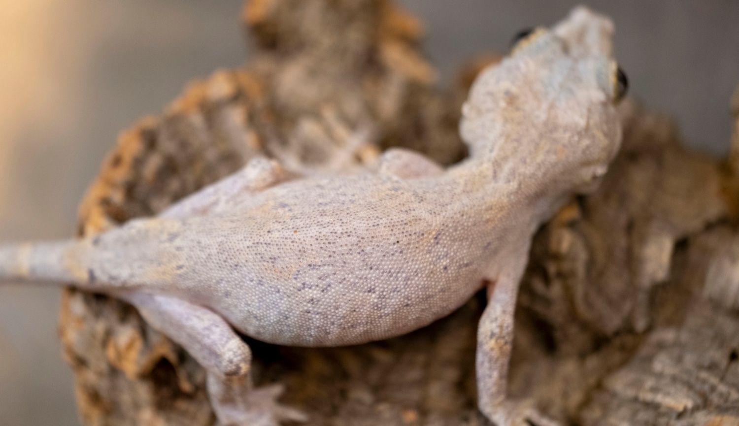
Many species have the ability to translocate the pigment inside chromatophores, resulting in an apparent change in colour. This process, known as physiological colour change, is most widely studied in melanophores, since melanin is the darkest and most visible pigment. In most species with a relatively thin dermis, the dermal melanophores tend to be flat and cover a large surface area. However, in animals with thick dermal layers, such as adult reptiles, dermal melanophores often form three-dimensional units with other chromatophores. These dermal chromatophore units (DCU) consist of an uppermost xanthophore or erythrophore layer, then an iridophore layer, and finally a basket-like melanophore layer with processes covering the iridophores.
Both types of dermal melanophores are important in physiological colour change. Flat dermal melanophores will often overlay other chromatophores so when the pigment is dispersed throughout the cell the skin appears dark. When the pigment is aggregated towards the centre of the cell, the pigments in other chromatophores are exposed to light and the skin takes on their hue. Similarly, after melanin aggregation in DCUs, the skin appears green through xanthophore (yellow) filtering of scattered light from the iridophore layer. On the dispersion of melanin, the light is no longer scattered and the skin appears dark. As the other biochromatic chomatophores are also capable of pigment translocation, animals with multiple chromatophore types can generate a spectacular array of skin colours by making good use of the divisional effect. [2]
Proven Lines
No known proven lines
Related Traits
No known related traits